Lower Leg Bone Diagram / Pelvic Girdle And Lower Limb Overview And Surface Anatomy Clinical Gate / Knee human anatomy function parts conditions 8 4 bones of the lower limb anatomy and physiology.
Lower Leg Bone Diagram / Pelvic Girdle And Lower Limb Overview And Surface Anatomy Clinical Gate / Knee human anatomy function parts conditions 8 4 bones of the lower limb anatomy and physiology.. The bones of the leg are the femur, tibia, fibula and patella. Long bone anatomy diagram 12 photos of the long bone anatomy diagram gross anatomy typical long bone diagram, long bone diagram quiz, long bone diagram unlabeled, long bone structure diagram, long. At the microscopic level, this hard outer. The bones involved in it, however, are only the femur and the tibia, although the. Name the 7 bones of the foot (not counting the phalanges).
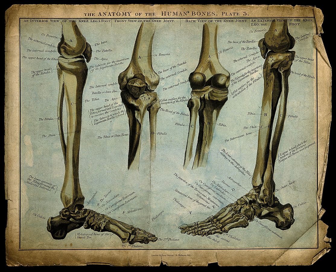
Posted on january 21, 2015 by admin. At the distal end of the femur, two rounded condyles meet the tibia and fibula bones of the lower leg to form the knee joint. Labeled diagram of long bone. Vector illustration with human skeleton scheme isolated on a white background. Master leg and knee anatomy using our topic page.
Ankle Fractures Broken Ankle Florida Orthopaedic Institute from www.floridaortho.com Master leg and knee anatomy using our topic page. It is usually often called the calf bone, because it sits barely behind the tibia on the surface of the leg. Your upper and lower leg are connected by a hinge joint. Health diagram bone skeleton leg knee science anchor chart human human body. While their parts are similar in general, their structure has been adapted to differing functions. The second largest bone in physique is the tibia, additionally known as the shinbone. Ankle human anatomy image function conditions more. Your leg bones are the longest and strongest bones in your body.
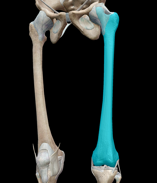
The femur, or thigh bone, is the largest, heaviest, and strongest bone in the human body.
The two bones beneath your knee that make up your shin are your tibia and fibula. 16 anterior muscles muscleorigininsertionfunction/action tibialis anteriortibiamedial cuneiform & 1 st metatarsal df and. The lower leg has a structure by two bones. While their parts are similar in general, their structure has been adapted to differing functions. The largest and most medial leg bone, forming both the knee and ankle joints. Download a free preview or high quality adobe illustrator ai, eps, pdf and high resolution jpeg versions. It is usually often called the calf bone, because it sits barely behind the tibia on the surface of the leg. The bones involved in it, however, are only the femur and the tibia, although the. Long bone anatomy diagram 12 photos of the long bone anatomy diagram gross anatomy typical long bone diagram, long bone diagram quiz, long bone diagram unlabeled, long bone structure diagram, long. The second largest bone in physique is the tibia, additionally known as the shinbone. The bones of the leg are the femur, tibia, fibula and patella. Your upper and lower leg are connected by a hinge joint. Ankle human anatomy image function conditions more.
The two arrows indicate where one of the bones of the leg (the tibia) is broken. Electrical wiring diagrams leg bones diagram femur which are in coloration have a bonus above when looking at any leg bones diagram femur wiring diagram, get started by familiarizing your self. The bones involved in it, however, are only the femur and the tibia, although the. Leg bone anatomy diagram diagram of human leg human anatomy. The artist's guide to the.
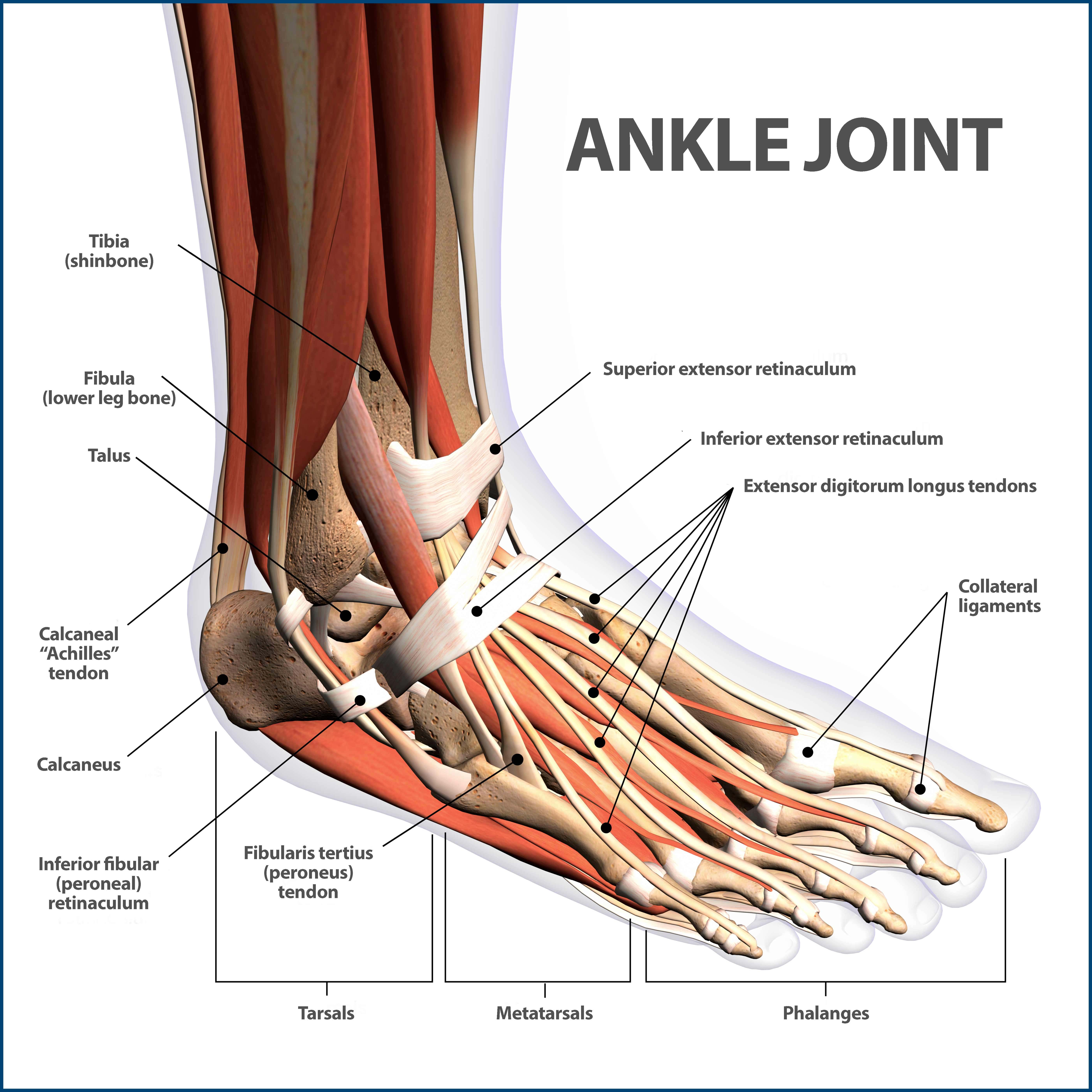
Bones Of The Lower Extremity Femur Tibia Fibula Phalanges And More from d3uigcfkiiww0g.cloudfront.net The foot bones shown in this diagram are the talus, navicular, cuneiform, cuboid, metatarsals and calcaneus. The bones involved in it, however, are only the femur and the tibia, although the. Related posts of bone anatomy lower leg. At the distal end of the femur, two rounded condyles meet the tibia and fibula bones of the lower leg to form the knee joint. At the microscopic level, this hard outer. Normal leg bones are relatively straight, but those affected by paget's disease are porous and figure 9. The bones of the leg are the femur, tibia, fibula and patella. Master leg and knee anatomy using our topic page.
By natalia kremenon january 21, 2021in wiring diagram231 views.
Vector illustration with human skeleton scheme isolated on a white background. It is the tibial joint surface or ceiling of the ankle mortise. Calcaneus, talus, navicular medial cuneiform, intermediate cuneiform, lateral cuneiform and cuboid. Ankle and foot bones and joints unit 4/12/18 lower leg: What is the weight bearing bone of the lower leg? Labeled diagram of long bone. Human skeleton long bones of arms and legs britannica. 16 anterior muscles muscleorigininsertionfunction/action tibialis anteriortibiamedial cuneiform & 1 st metatarsal df and. Download a free preview or high quality adobe illustrator ai, eps, pdf and high resolution jpeg versions. The humerus and the femur are corresponding bones of the arms and legs, respectively. Bones of the leg and foot, lower leg bone anatomy, leg bones anatomy, leg muscles, leg bones diagram, leg bone structure, leg anatomy muscles, parts of the lower leg. By natalia kremenon january 21, 2021in wiring diagram231 views. Ankle human anatomy image function conditions more.
Calcaneus, talus, navicular medial cuneiform, intermediate cuneiform, lateral cuneiform and cuboid. Labeled diagram of long bone. 2006 kia optima belt diagram. Your leg bones are the longest and strongest bones in your body. Bones of the lower limb anatomy and physiology i.
3d Skeletal System 5 Cool Facts About The Femur from www.visiblebody.com When you stand or walk, all the weight of your upper body rests on them. Anterior view with primary bones names. At the distal end of the femur, two rounded condyles meet the tibia and fibula bones of the lower leg to form the knee joint. 2006 kia optima belt diagram. The two bones beneath your knee that make up your shin are your tibia and fibula. Cheek bone (zygoma) upper jaw (maxilla). Distal end of tibia that forms the medial ankle 15 posterior muscle diagram. Vector illustration with human skeleton scheme isolated on a white background.
2006 kia optima belt diagram.
Health diagram bone skeleton leg knee science anchor chart human human body. Ankle and foot bones and joints unit 4/12/18 lower leg: Normal leg bones are relatively straight, but those affected by paget's disease are porous and figure 9. What is the weight bearing bone of the lower leg? Download a free preview or high quality adobe illustrator ai, eps, pdf and high resolution jpeg versions. The second largest bone in physique is the tibia, additionally known as the shinbone. The knee is a strong but flexible hinge joint. Posted on april 18, 2019april 18, 2019. Long bone anatomy diagram 12 photos of the long bone anatomy diagram gross anatomy typical long bone diagram, long bone diagram quiz, long bone diagram unlabeled, long bone structure diagram, long. Your leg bones are the longest and strongest bones in your body. License image the bones of the leg are the femur, tibia, fibula the foot bones shown in this diagram are the talus, navicular, cuneiform, cuboid, metatarsals and calcaneus. 16 anterior muscles muscleorigininsertionfunction/action tibialis anteriortibiamedial cuneiform & 1 st metatarsal df and. The bones of the leg are the femur, tibia, fibula and patella.
The human leg, in the general word sense, is the entire lower limb of the human body, including the foot, thigh and even the hip or gluteal region leg bone diagram. Master leg and knee anatomy using our topic page.


0 Komentar